
In 2001, a research team at the Hebrew University of Jerusalem witnessed a remarkable scene in the laboratory. When they took an arm of a living Octopus vulgaris (the common octopus), severed its connection to the central brain, and gently stimulated it, the arm reproduced a perfect reaching motion entirely on its own. Even without any command from the brain, the arm controlled its own muscles and exhibited kinematics almost identical to those of an intact arm. An entire motor program was embedded within the arm itself (Sumbre et al. 2001, Science).
This discovery distills the core design principle of the octopus nervous system. The octopus is not an animal in which “a central command headquarters controls everything.” Instead, it delegates high-level goals to the arms, and the arms themselves compute the detailed behavior independently. By following how this structure works — and why a color-blind animal can pull off color camouflage so refined that it is hard to distinguish from the background — we can see the principles of this finely designed distributed intelligence come into view.
Nine brains? More precisely, a “1+8 distributed structure”
The phrase “the octopus has nine brains” is widely used, but it is a metaphor rather than a biological fact. The octopus has one central brain. It is a mass of nervous tissue that encircles the esophagus like a ring, divided into upper and lower portions and composed of roughly 40 interconnected lobes. And in each of the 8 arms runs an axial nerve cord (ANC) and the ganglia within it. These arm ganglia act as semi-independent processing centers, but they are not anatomically separate “brains” (Hochner 2012, Current Biology).

Jouni Kuisma, DiveCodex · CC BY-SA 4.0 · Wikimedia Commons · Source
The numbers are even more striking. The octopus has a total of about 500 million neurons, on par with a vertebrate such as a dog, and it is the largest nervous system among invertebrates. Yet only about 40–45 million of these (roughly 8% of the total) are in the central brain. The optic lobes hold about 120–180 million, and the remaining roughly 330 million (about two-thirds of the total) are distributed across the arms. The connection linking the brain and the arm nervous system consists of only about 32,000 efferent and about 140,000 afferent nerve fibers (Zullo et al. 2019, Journal of Comparative Physiology A; species: Octopus vulgaris). This sparse connection is the physical basis for peripheral autonomous processing.
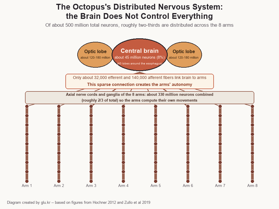
Diagram · created by glu.kr
What does this structure mean? The 8 arms are “infinite degrees of freedom” structures with neither a backbone nor joints. For the brain to micro-control hundreds of millions of muscle fibers in real time, the computational load would become astronomical. The octopus’s solution was “delegation.” The brain conveys a high-level goal such as “catch the prey,” and the neural circuits inside the arm autonomously compute the specific muscle patterns. In 2025, a study on Octopus bimaculoides confirmed at the molecular level that the arm ANC has a segmented structure corresponding to each individual sucker. This means a sucker-specific map (suckerotopy) is formed, in which a dedicated neural module is assigned to each sucker (Olson et al. 2025, Nature Communications).
As a result, the octopus is short-lived. Most species live 1–2 years and breed only once in their lifetime before dying (semelparity). It is astonishing that this complex nervous system operates over such a brief life span.
Three hearts, blue blood — the physiology that sustains the distributed nervous system
To maintain a distributed nervous system, the energy supply must be stable. The octopus’s circulatory system is designed for this task. It has three hearts. Two branchial hearts each send blood to a gill to supply oxygen, and one systemic heart receives the oxygenated blood and sends it throughout the body.
This three-heart structure is necessary because hemocyanin, the molecule that carries oxygen in the blood, has low efficiency. Hemocyanin is a copper (Cu)-based protein, and when it binds oxygen it takes on a blue color — which is why the octopus’s blood is blue. Its oxygen-carrying efficiency is only about one-quarter that of iron-based hemoglobin (red blood). The three hearts compensate for this low efficiency.
Paradoxically, this “inefficient” molecule becomes a strength in cold-water environments. The Antarctic octopus (Pareledone charcoti) raises its hemocyanin concentration by more than 40% compared with warm-water species, and is functionally tuned to maintain oxygen-release efficiency even at sub-zero water temperatures (Oellermann et al. 2015, Frontiers in Zoology). It is an oxygen carrier specialized for cold, low-oxygen environments.
Suckers: 1,920 combined touch-and-taste sensors
Octopus vulgaris arranges about 240 suckers per arm in two rows, totaling about 1,920 across its 8 arms. These suckers are not simple suction organs.

Zi X. Ou · CC0 · Wikimedia Commons · Source
In 2020, a Harvard University research team discovered two types of sensory cells in the suckers of the California two-spot octopus (Octopus bimaculoides). The chemosensory cells express a new family of chemotactile receptors (CRs) to detect terpenoid molecules from prey, and the mechanosensory cells transmit instantaneous touch via NompC receptors. A single sucker simultaneously touches and tastes (touch-taste) (van Giesen et al. 2020, Cell). There are about 30 million receptor neurons per arm, reaching about 240 million across the 8 arms.
This abundant peripheral sensory information is processed first in the arm ganglia. The advantage of the distributed nervous system appears here as well. Rather than touch information having to make a round trip to the brain, local judgments are made in the arm itself.
Color-blind yet color-camouflaging — an unsolved paradox
The octopus’s most striking ability is camouflage so refined that it is hard to distinguish from the background. It replicates the color and texture of a reef within hundreds of milliseconds. Yet the octopus is a monochromat, possessing only a single type of photoreceptor in its retina (Stubbs & Stubbs 2016, PNAS). How does an animal that cannot distinguish color carry out color camouflage?
This paradox has not yet been fully resolved. Two hypotheses have been proposed.
Hypothesis A — pupillary chromatic aberration: Stubbs & Stubbs (2016, PNAS), in a modeling study covering three cephalopod groups — octopus, cuttlefish, and squid — proposed that the octopus’s distinctive dumbbell-shaped pupil maximizes the lens’s wavelength-dependent refraction differences (chromatic aberration). The idea is that even with only a single-color receptor, the animal can infer color information by reading “at which wavelength the image is focused most sharply.” However, this is a hypothesis based on computer simulation and has not been fully verified by behavioral experiments.
Hypothesis B — skin opsins: Ramirez & Oakley (2015, Journal of Experimental Biology) confirmed the expression of a protein of the same family as the eye’s photoreceptor protein (r-opsin) in the skin of Octopus bimaculoides. When blue light (λmax ≈ 480 nm) was shone on a piece of skin, a phenomenon was observed in which the chromatophores expanded autonomously without input from the brain or eyes (“light-activated chromatophore expansion,” LACE). This raises the possibility that the entire skin acts as a low-resolution light detector to assist color camouflage. This, too, remains unverified as to how much it actually contributes in natural camouflage.
The two hypotheses may not be mutually exclusive, and neither is an established fact — both are “proposed mechanisms.” The paradox of color blindness and color camouflage is an ongoing research topic.
Chromatophore, iridophore, leucophore — the three-layer structure of camouflage
Separately from how it senses color, the mechanism by which the skin produces color is well understood. The octopus skin coordinates three layers from the surface down to the deep layer.
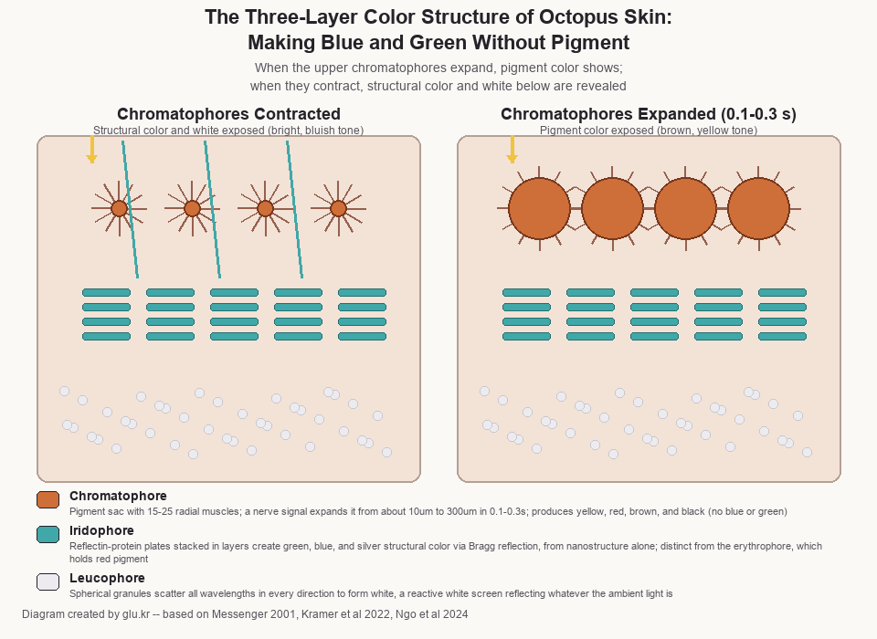
Diagram · created by glu.kr
Surface layer — chromatophore: a structure in which 15–25 radial muscles surround a pigment sac (cytoelastic sacculus). When a nerve signal is delivered directly to the muscles, the diameter expands from about 10 μm to about 300 μm — nearly 30-fold — within 0.1–0.3 s (Messenger 2001, Biol. Rev.; subjects: cephalopods including Octopus vulgaris). The pigment colors are in the yellow, red, orange, brown, and black range. Importantly, chromatophores cannot produce blue and green. These wavelengths are handled by the structural-color cells in the layers below.
This color change has a very high energy cost. According to a 2024 study on Octopus rubescens (Ngo et al., PNAS), the amount of oxygen consumed when fully activating the chromatophores reaches about 92% of the resting metabolic rate of a 100 g octopus. Maintaining camouflage is, for the octopus, an activity that consumes nearly twice the energy.
Middle layer — iridophore (structural-color reflection): inside the cell, platelet structures made of the reflective protein reflectin are stacked in multiple layers. The spacing between the plates selectively reflects light of specific wavelengths by Bragg reflection, producing structural colors in the green, blue, and silver range. Color is generated by nanostructure alone, without any pigment. Unlike in squid, the octopus’s iridophores are generally believed to have fixed plate spacing, serving to statically reflect specific wavelengths (Kramer et al. 2022, Int. J. Mol. Sci.). Note: the iridophore is a structural-color cell and is entirely different from the erythrophore, which holds red pigment.
Deep layer — leucophore: small spherical granules scatter all visible wavelengths in every direction, producing a white appearance. As a “reactive white screen” that reflects whatever wavelength is in the ambient lighting, it contributes to adjusting contrast and brightness.
When the chromatophores expand, the two layers beneath are hidden and only the pigment color is seen; when the chromatophores contract, the structural-color and white layers are revealed. From the moment vision processes a stimulus to the moment a pattern is formed on the skin, a three-stage neural hierarchy running from the brain to the skin (optic lobe → lateral basal lobe → chromatophore lobe) transmits the signal within hundreds of milliseconds.
In addition, skin papillae can convert the two-dimensional flat skin into three-dimensional relief. The papillae are muscular hydrostats that maintain their shape with muscle alone, without any skeleton (studies of Octopus vulgaris and O. bimaculoides, Allen et al. 2013). This function adds texture camouflage to color camouflage, disrupting the outline.
Selective background matching — it does not average the whole
A study of Octopus cyanea and O. vulgaris (Josef et al. 2012, PLOS ONE) confirmed an intriguing fact. The octopus does not match its color by averaging the entire environment in which it sits. Instead, it selectively extracts only the color and texture of a specific immediately adjacent object — a particular coral fragment, a particular rock patch — and matches its skin pattern to it. Of 11 individuals, 10 showed a match to the immediately adjacent landmark pattern. This is local feature extraction, not a global average.
Behavioral abilities — tool use, mimicry, observational learning
The behavioral abilities produced by the distributed nervous system and refined skin control are impressive.

q phia · CC BY 2.0 · Wikimedia Commons · Source
Tool use: In waters off Indonesia, a veined octopus (Amphioctopus marginatus) was observed carrying a coconut shell split in half for more than 20 m and later assembling it into a shelter. During transport the shell provides no protection, and the octopus has to perform an awkward “stilt-walking” gait. This is “deferred utility” tool use, in which a present cost is borne for a future benefit — the first official report in an invertebrate (Finn et al. 2009, Current Biology).

Rickard Zerpe · CC BY-SA 2.0 · Wikimedia Commons · Source
Mimicry: The mimic octopus (Thaumoctopus mimicus) has been reported to change not only its skin color and texture but also the posture, shape, and mode of movement of its arms to imitate various marine animals such as sea snakes, lionfish, and soles (Norman et al. 2001, Proc. R. Soc. B). There are also field observations that when it encounters a damselfish that harasses it, it transforms into a sea snake — the fish’s natural enemy — to drive it off. Dynamic mimicry, switching the target of imitation depending on the situation, is so far most prominently known in this species (species description: Norman & Hochberg 2005, Molluscan Research).
Observational learning: In an experiment on Octopus vulgaris (Fiorito & Scotto 1992, Science), an octopus that observed a demonstrator individual select one of two colored spheres four times consistently chose the same sphere as the demonstrator in a subsequent independent test. It was the first demonstrated case of observational learning (social learning) in an invertebrate. The result is all the more remarkable given that the octopus is fundamentally a solitary animal.
Its color changes even during sleep
Evidence that chromatophore control operates independently of conscious judgment came from sleep research. In a 2023 study on Octopus laqueus (Pophale et al., Nature), an “active sleep” state was identified in which, at intervals of roughly one hour between bouts of quiet sleep, the skin color changed rapidly for tens of seconds at a time to the same level as in the waking state. Eye movements and sucker contractions also accompanied it. The possibility was raised that this is a neural pattern that convergently evolved with mammalian REM sleep. Although the research team stated explicitly that there is no way to confirm whether the octopus dreams, the very fact that chromatophore control can operate independently of central consciousness adds a new dimension to the discussion of the distributed nervous system.
Refinement without central control — the meaning of the design principle
What the octopus shows is an alternative to “centralized design.” Even without the brain dictating every detail, local algorithms embedded in the arms coordinate movement, sensation, and response. Chromatophores, iridophores, and leucophores coordinate across three layers to produce structural color without any pigment signal, and it is even proposed that the skin itself may sense light and respond.

Julia Markey · CC BY-SA 4.0 · Wikimedia Commons · Source
This architecture is also a biological model frequently referenced today in research on soft robotics and distributed control systems. The principle that each module produces refined overall behavior from local information without any single blueprint — the octopus is living proof that this is biologically feasible.
References
- Sumbre G et al. (2001). Control of octopus arm extension by a peripheral motor program. Science 293: 1845–1848.
- Zullo L et al. (2019). Motor control pathways in the nervous system of Octopus vulgaris arm. J Comp Physiol A.
- Hochner B (2012). An Embodied View of Octopus Neurobiology. Current Biology 22: R887–R892.
- van Giesen L et al. (2020). Molecular Basis of Chemotactile Sensation in Octopus. Cell 183: 594–604.
- Stubbs AL & Stubbs CW (2016). Spectral discrimination in color blind animals via chromatic aberration and pupil shape. PNAS 113: 8206–8211.
- Ramirez MD & Oakley TH (2015). Eye-independent, light-activated chromatophore expansion and expression of phototransduction genes in the skin of Octopus bimaculoides. J Exp Biol 218: 1513–1520.
- Finn JK et al. (2009). Defensive tool use in a coconut-carrying octopus. Current Biology 19: R1069–R1070.
- Ngo V et al. (2024). Metabolic cost of chromatophore activation in Octopus rubescens. PNAS.
- Pophale A et al. (2023). Wake-like skin patterning and neural activity during octopus sleep. Nature.
- Albertin CB et al. (2015). The octopus genome and the evolution of cephalopod neural and morphological novelties. Nature 524: 220–224.
- Olson CL et al. (2025). Segmented organization of the octopus arm nervous system. Nature Communications (DOI: 10.1038/s41467-025-56558-7).
- Josef N et al. (2012). Camouflage during movement in the day octopus (Octopus cyanea). PLOS ONE 7(5): e37579.
- Messenger JB (2001). Cephalopod chromatophores: neurobiology and natural history. Biological Reviews 76: 473–528.
- Kramer RM et al. (2022). Reflectin and structural coloration in cephalopods. Int. J. Mol. Sci. 23: 11546.
- Allen JJ et al. (2013). Cephalopod skin papillae and three-dimensional camouflage. Journal of Morphology 274: 175–190.
- Fiorito G & Scotto P (1992). Observational learning in Octopus vulgaris. Science 256: 545–547.
- Norman MD, Finn J & Tregenza T (2001). Dynamic mimicry in an Indo–Malayan octopus. Proc. R. Soc. B 268: 1755–1758.
- Oellermann M et al. (2015). Blue blood on ice: blood oxygen transport in an Antarctic octopod. Frontiers in Zoology 12: 6.